A lines
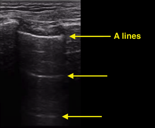
"A lines" are one of the normal artifacts that are seen in lung ultrasound.
This is what they look like:

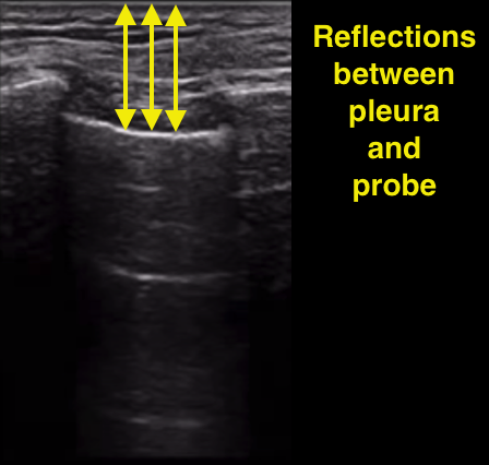
They are caused by something called reverberations. A reverberation occurs when the ultrasound signal bounces back and forth in between 2 structures and every time is bounces "back" to the probe, it sends a signal to the ultrasound machine. In the case of A lines, the ultrasound signal bounces between the parietal pleura and the probe itself.

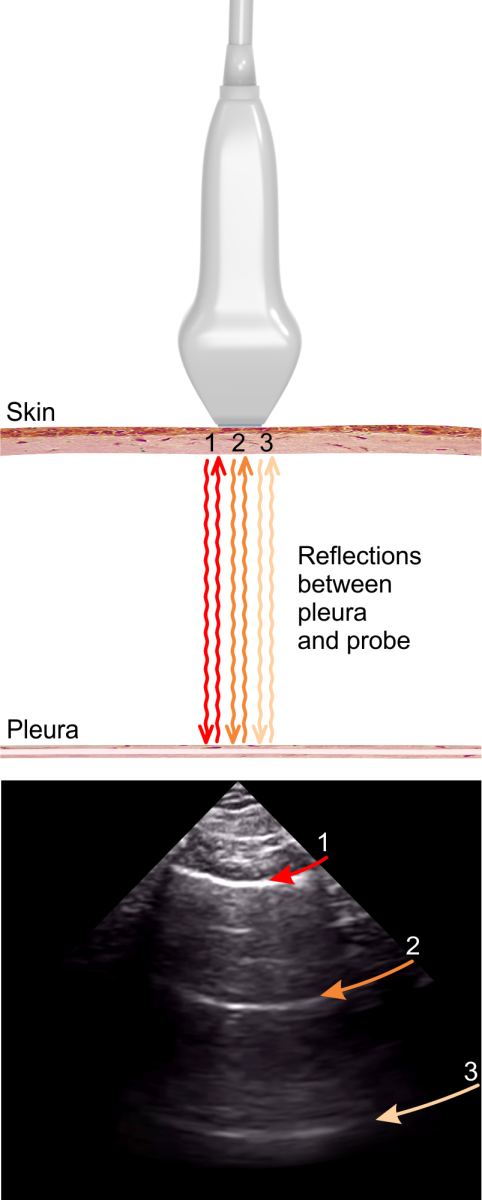
Shown a little differently below is each successive "bounce" between the pleura and transducer using different colored arrows. The ultrasound machine uses time to measure depth: the further away something is, the more time it will take for the signal to return to the probe.
In the image below, the first reverb is red, followed by orange and then yellow. See how the first reverberation (red) is closer to the top of the ultrasound image than the latter reverberations?

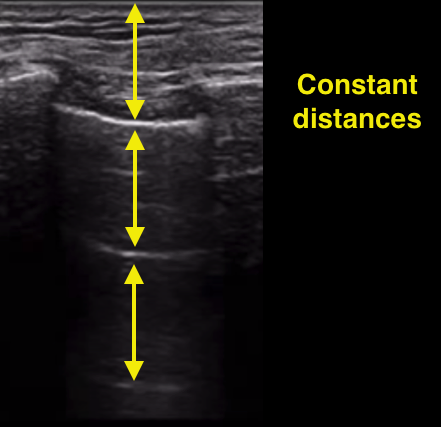
Each bounce travels the same distance. As a result, the timing in between each bounce is constant. Therefore, constant time intervals between each reverberation results in constant distances between each A line:

Here is an important detail: in order for these reverberations to occur, air must be immediately below the parietal pleura. Remember that the parietal pleura is part of the chest wall, while the visceral pleura is the outer lining of the lung itself.
A normal air filled lung will create A lines, but a pneumothorax will also create A lines.
If the lung is full of fluid or pus (as in heart failure, ARDS or pneumonia), then A lines will not occur.
Summary of A lines:
- A lines are an artifact caused by reverberation
- A lines are a normal finding
- A lines require air to be present immediately below the parietal pleura
- A lines are constant distances apart
- A lines can exist with pneumothorax